Anatomical Illustrations
Commission and independent work
2019- current
2019- current
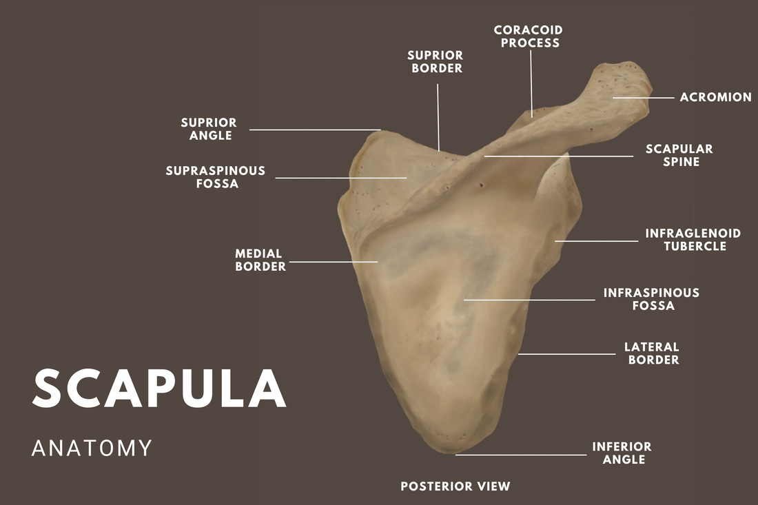
Scapula Anatomy
Photoshop
2022
Photoshop
2022
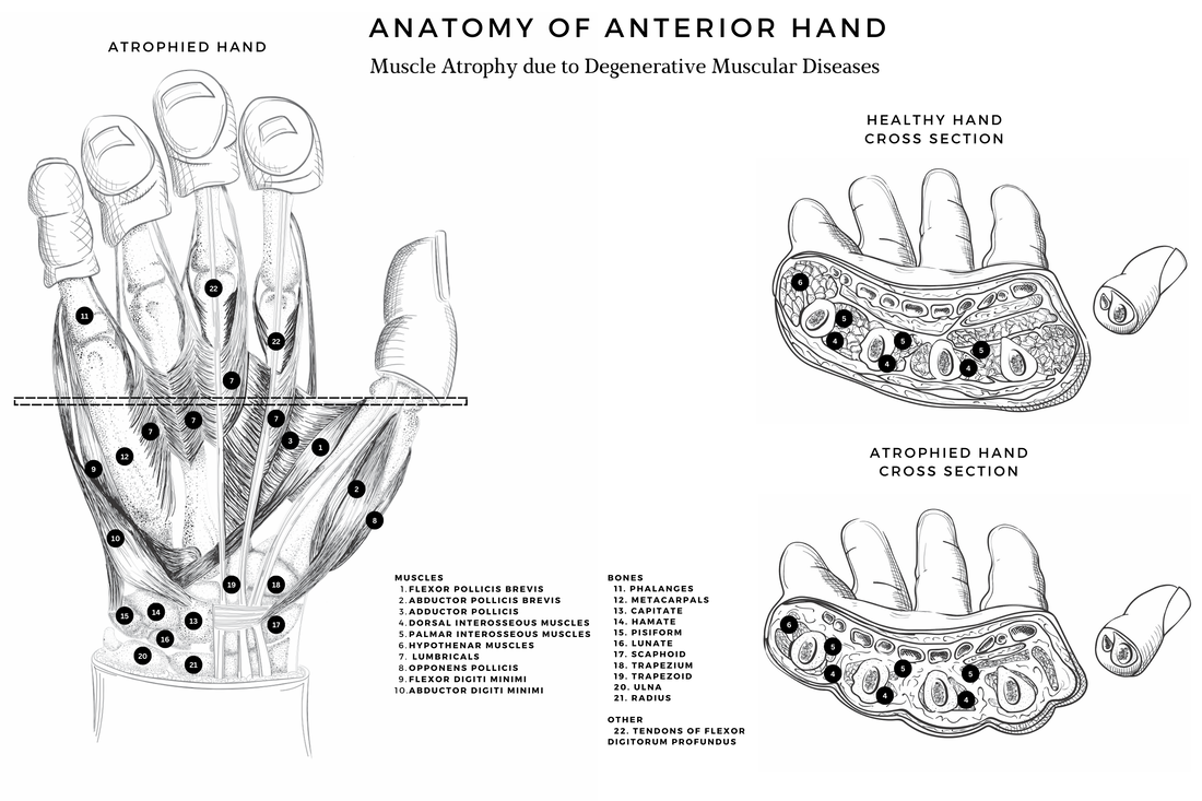
Muscular Atrophy Anatomy
Photoshop
2022
Photoshop
2022
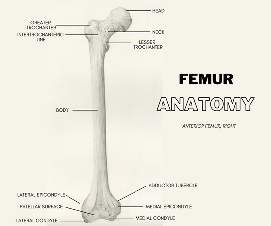
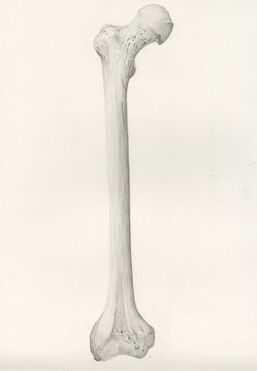
Femur Anatomy
Graphite, digitally labeled
2022
Graphite, digitally labeled
2022
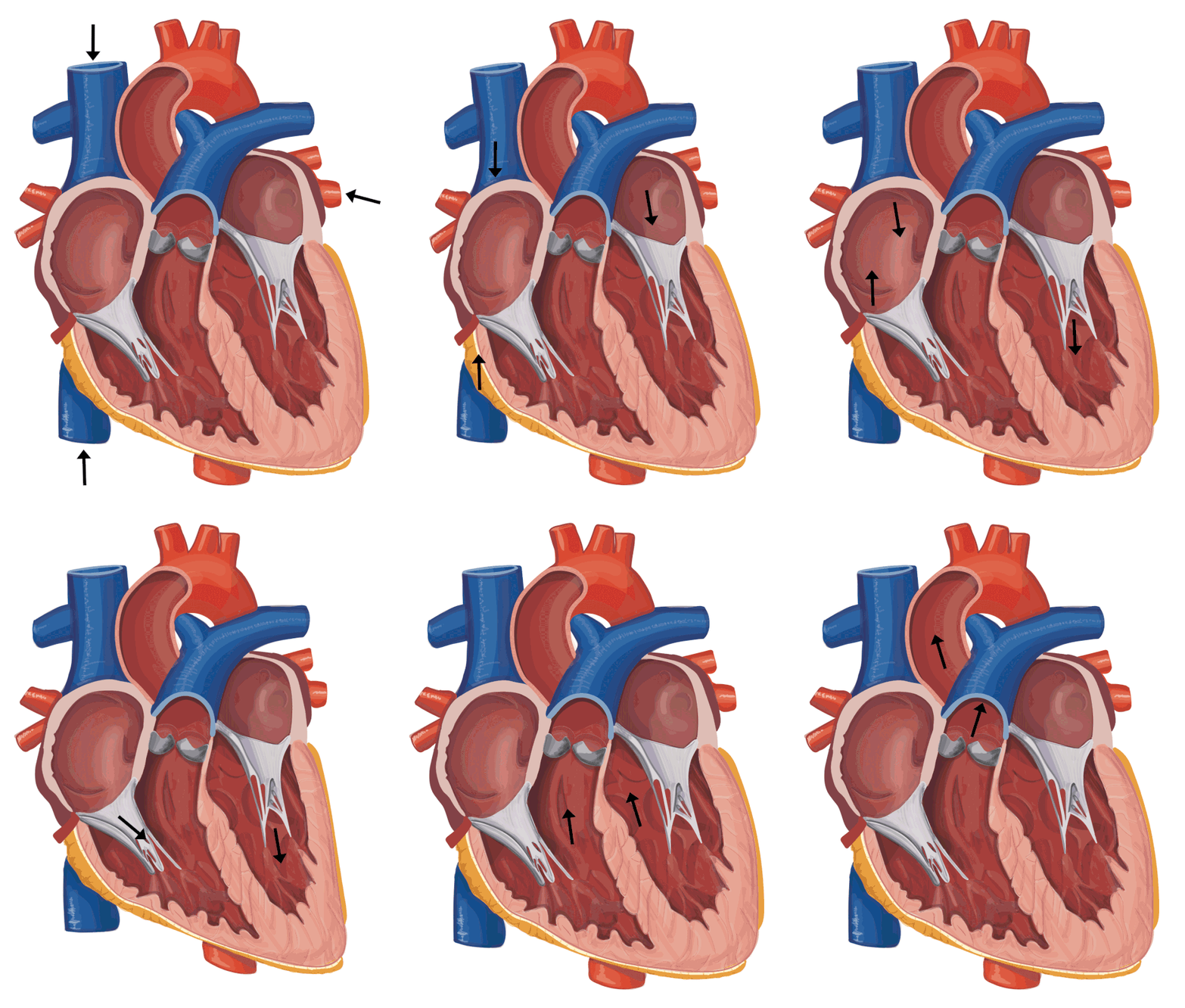
Normal Heart Beat Animation
Photoshop, Illustrator
2021
Photoshop, Illustrator
2021
This animation is part of a work in progress which is a series of animations and illustrations showing normal heart functions compared to hearts with congenital diseases
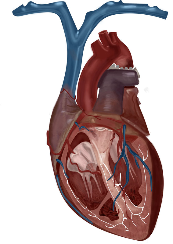
Human Heart Cross Section
2019
Adobe Illustrator
2019
Adobe Illustrator
Hernia Anatomy project
2020
Adobe Illustrator
2020
Adobe Illustrator
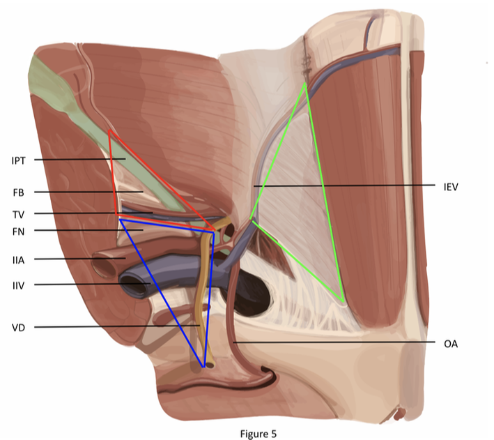
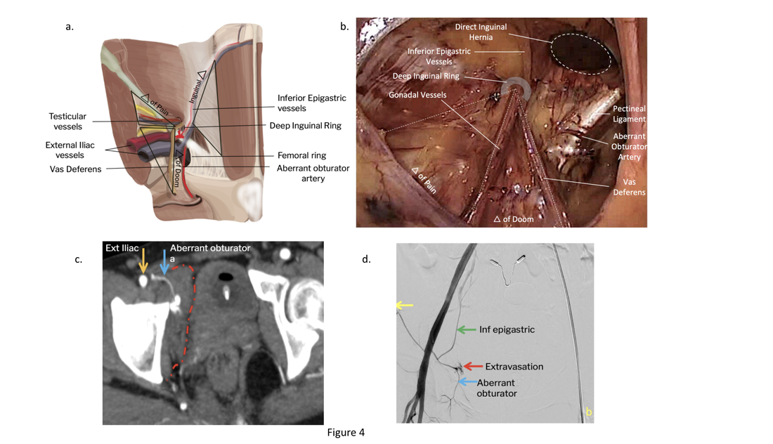
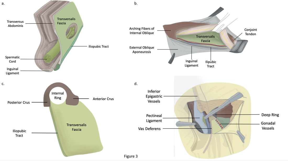
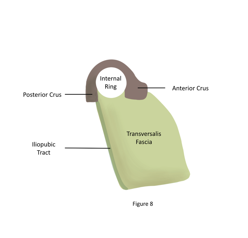
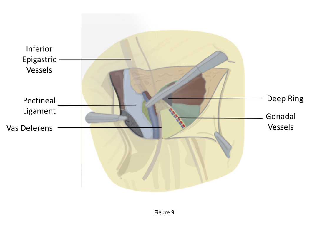
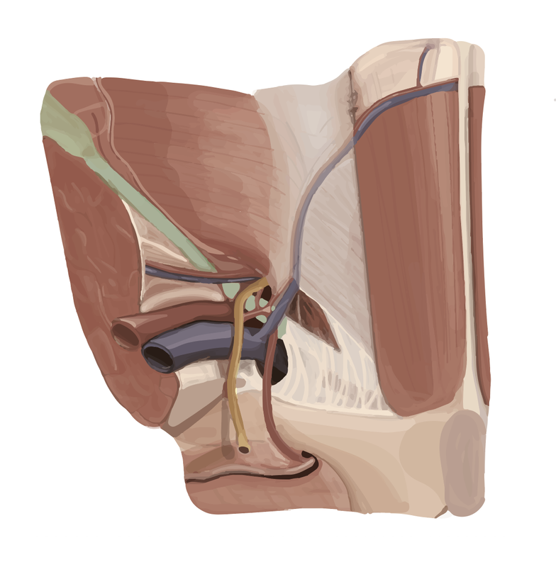
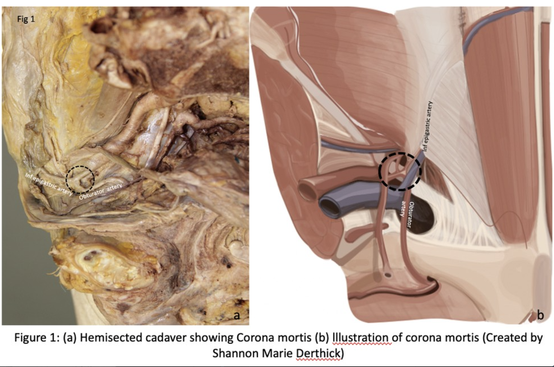
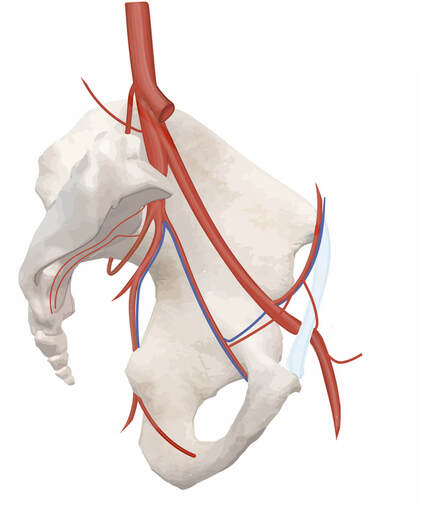
Hernia repair is a relatively common surgery. In recent years, there have been major changes in the laparoscopic surgery and use of mesh prosthetics. Th information needed for understanding the inguinal region and what is taught in medical schools is outdated compared to the procedure. Some of the major anatomical landmarks of the inguinal region that are traditionally taught to medical students for example are the median, medial, and lateral umbilical folds, inferior epigastric vessels, inguinal triangle, and the deep and superficial inguinal rings. The anatomical essentials must be updated to match the current anatomical language and terms used during surgery. The triangle of doom refers to a triangular area bound by the vas deferens, the testicular vessels and the peritoneal fold and is an important landmark during laparoscopic inguinal hernia repair. In the boundaries of this area are also the external iliac artery and vein.
Figure 5. Illustration of posterior view of inguinal region. Iliopubic Tract (IPT), Femoral Branch of the Genitofemoral Nerve (FB), Testicular Vessels (TV), Femoral Nerve (FN), Internal Iliac Artery (IIA), Internal Iliac Vein (IIV), Vas Deferens (VD), Inferior Epigastric Vessels (IEV), Obturator Artery (OA), Triangle of Pain (Red), Triangle of Doom (Blue), Inguinal Triangle (Green)
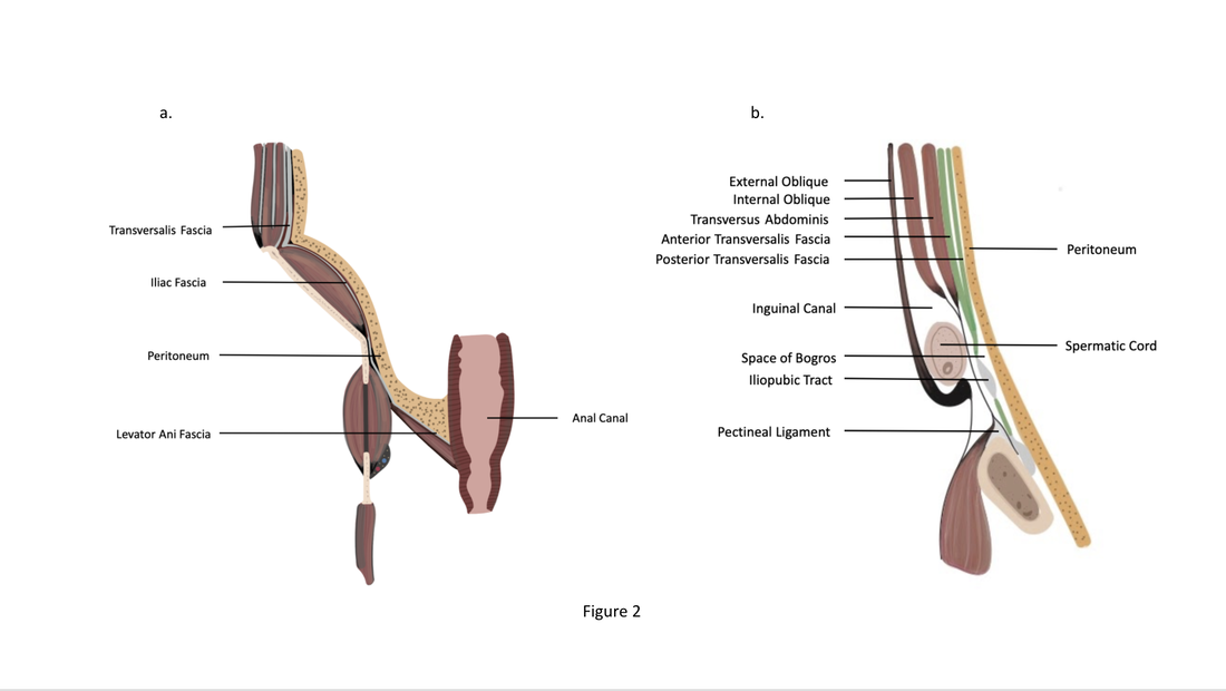
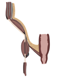
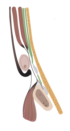
Figure 4. Midline sagittal view of structures forming the anterior abdominal/inguinal wall. Anterior Lamina of Transversalis Fascia (Anterio TF), Posterior Lamina of Transversalis Fascia (Posterior TF)
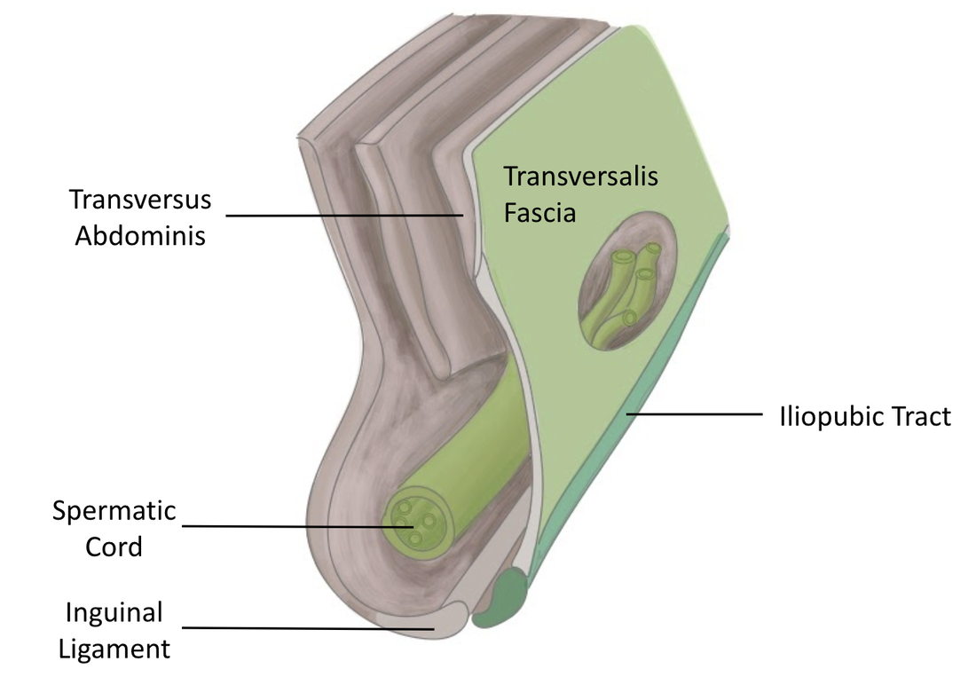
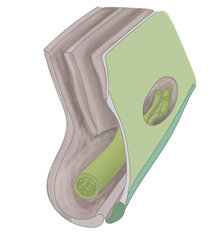
Figure 3. Illustration of the continuity of abdomino-inguino-pelvic fascia from a frontal view.
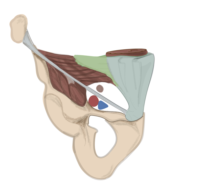
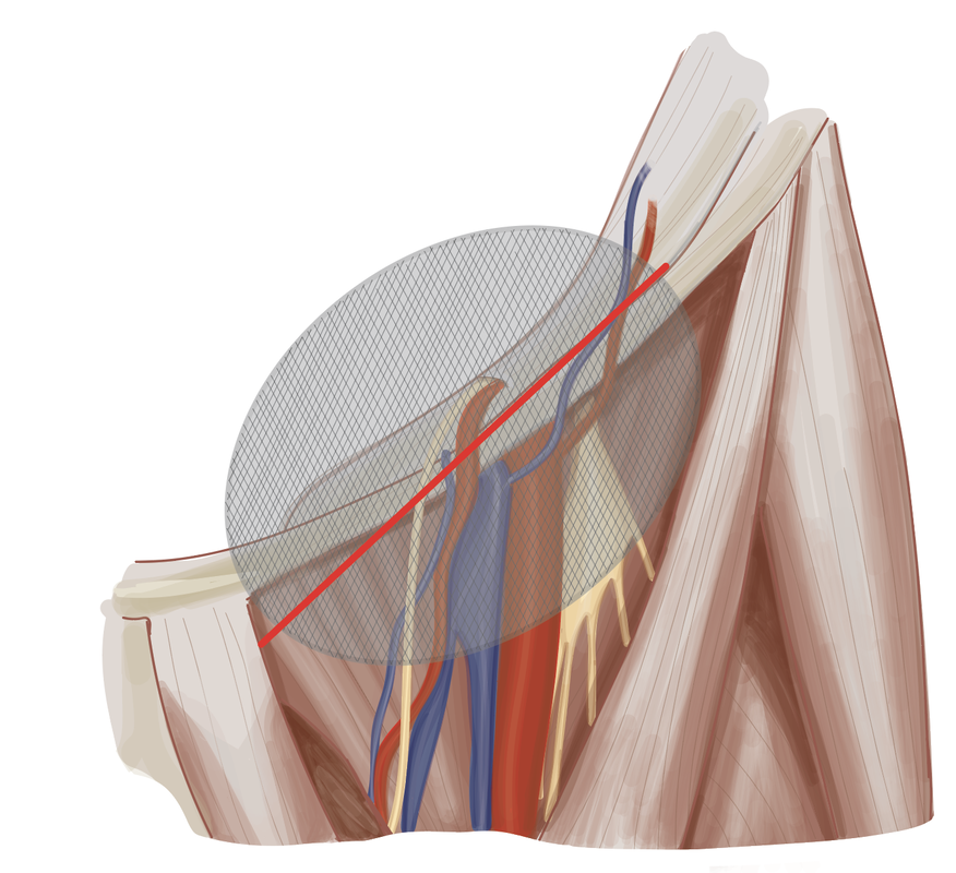
Figure 2. Illustration of Myopectineal Orifice and surrounding structures. The red enclosure represents the suprainguinal space and the blue enclosure represents the infrainguinal space
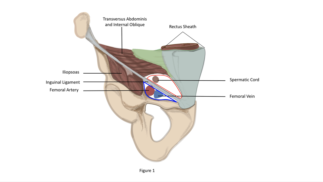
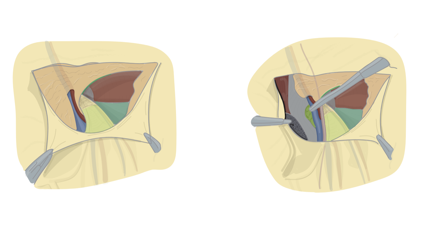
Figure 1. Operational Image highlighting traditional inguinal anatomic features. Median Umbilical Fold (Median UF), Medial Umbilical Fold (Medial UF), Inferior Epigastric vessels (IEV), Deep Inguinal Ring
Posters presented at AAA
2021
2021
These posters are the final educational product of the hernia illustrations
www.oakland.edu/Assets/Oakland/medicine/graphics/faculty/Medical-Education-Week/2021%20MedEdWeek%20-%20EdInnovationPosters.pdf
www.oakland.edu/Assets/Oakland/medicine/graphics/faculty/Medical-Education-Week/2021%20MedEdWeek%20-%20EdInnovationPosters.pdf
Unlabeled images
Human Eye
2019
Adobe illustrator
2019
Adobe illustrator
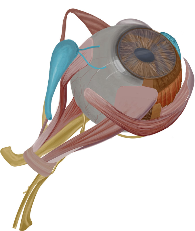
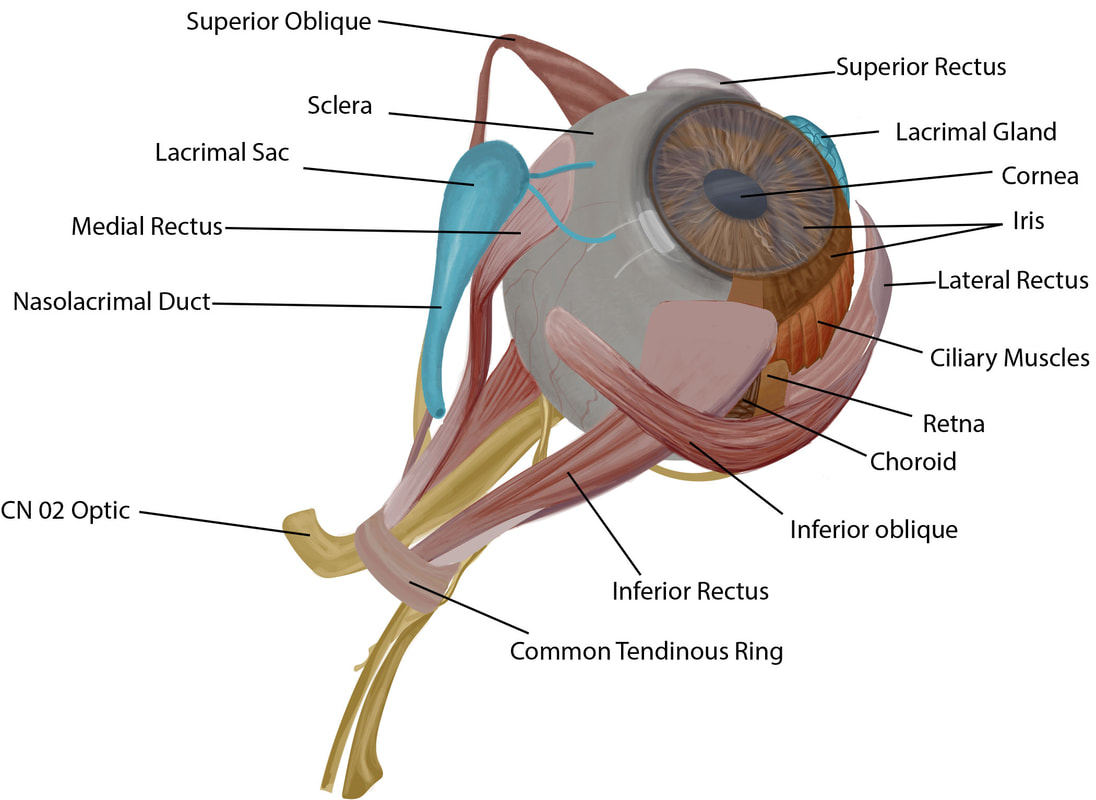
An independent project to learn the different parts of a human eye
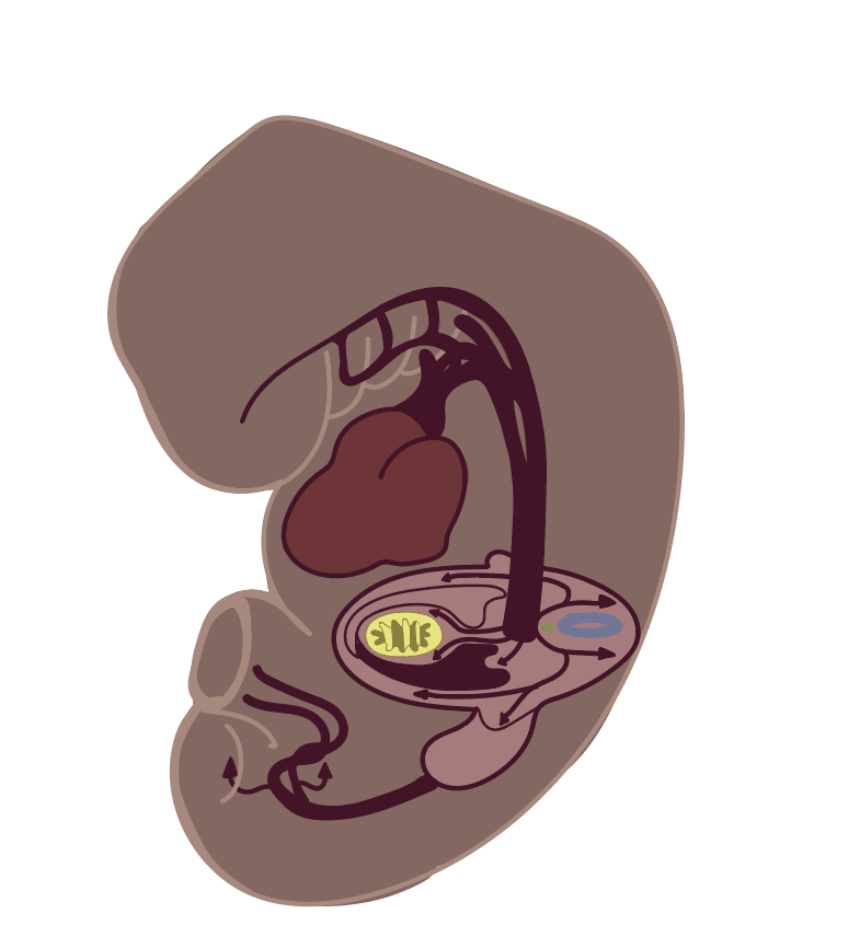
Embryo Project (unlabeled)
2021
Adobe Illustrator
2021
Adobe Illustrator
Nuraptive Therapeutics project
2019
Adobe Illustrator
2019
Adobe Illustrator
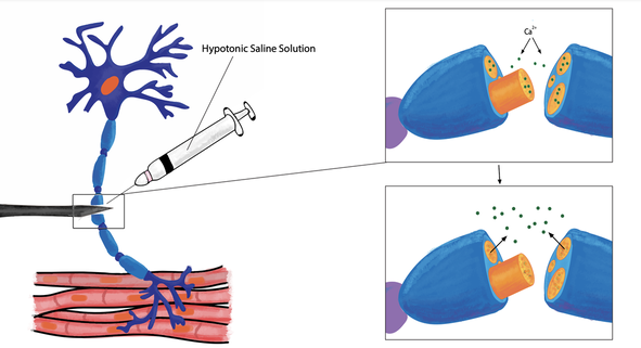
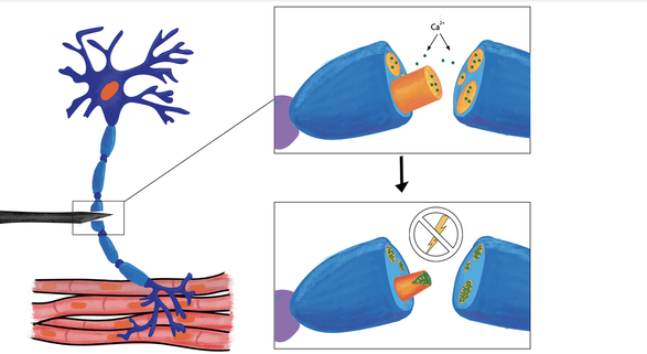
A serum developed by a biotechnology start-up can help establish electric charges within heavily damaged or severed nerve cells which leads to restoration of the nerve and regaining function.
STIMple Solutions project manual illustrations
2019
Adobe Illustrator
2019
Adobe Illustrator
|
|
|

